
I received the opportunity to work at the mechanobiology laboratory under the guidance of professor Dr Namrata Gundiah in the department of mechanical engineering.
The aim of my research project was to identify the effects of TGF beta and TGF beta inhibitor on the mechanical properties of MDAMB 231 invasive breast cancer cells and non invasive MCF-7 non invasive breast cancer cells. We aimed to study the morphological variation in the breast cancer cells following treatment with tgfb and tgfb inhibitor at time intervals of 24 and 48 hours and also to study the effects of shear stress on the deadhesion of these cells from the substrate following treatments with TGF beta and TGF beta inhibitors for 24 hours.
Since the laboratory primarily works on experiments involving traction force microscopy I also received an introduction into the protocol involved in the setting up of such an experiment.
Traction force microscopy is a technique that involves determining the tractions on the surface of a biological cells by measuring the displacements in the surrounding media which is detected by the displacements of microscopic fluorescent beads that are embedded in a uniplanar manner on the surface of a biocompatible gel atop which the cells are grown. The traction forces exerted by the cells cause the beads to move slightly and when the cells are washed off by a detergent solution the tension exerted by the cellular tractions get relieved causing the beads to slip back into their original positions. Measuring the displacement between the two positions of the beads surrounding a particular cell gives us an estimate of the cellular traction forces exerted by a particular cell.
In order to set up a traction experiment I learnt how to treat one set of square cover slips with 3- amino-propyl triethoxy silane followed by two washes with DI water and incubated them with 0.5% glutaraldehyde solution for 30 minutes at room temperature. Another set of circular cover slips were treated with bovine serum albumin in a humid chamber for 45 minutes. These circular cover slips were then spin coated (500 rpm, 2 minutes) with 20 micro litre of 2% fluorescent bead solution to ensure uniform distribution of the beads on the cover slip. Then a solution for 10kpa gel was prepared using N,N methylene bisacrylamide (40%wt/vol) and acrylamide (2% wt/vol). 200 micro litre of this solution was mixed with 2 micro litre of APS and 0.2 micro litres of TEMED(neurotoxic) and 30 micro litre of the mixture was pipetted onto the glutaraldehyde treated square coverslips and the bead coated circular coverslips were placed atop the droplet of solution so as to allow a thin layer of 10kpa stiffness gel to polymerise between the two coverslips.
The circular coverslips are then removed using a scalpel which causes the beads to be retained in the top layers of the gel on the square coverslips. These square coverslips are then carefully attached to punch Petri dishes using vacuum grease to create an air tight seal and the gels are kept moist using PBS solution. The gels are then treated with 200 microlitres of a solution of sulpho SANPAH heterobifunctional linker under 365 nm UV radiation for ten minutes. This process is repeated once following which 100 microlitres of rat tail collagen 1 is pipetted onto the surface and treated in the incubator for 45 minutes. The cells are then seeded onto these Petri dishes. The dishes are visualised under the epifluorescence microscope to ascertain the position of beads with the cells attached (stressed configuration) and following removal of the cells with a detergent solution which causes the cellular traction forces to relax and the beads to attain their original position (relaxed configuration) and the displacements and thereby the traction forces exerted by the cells are quantified using MATLAB software. [1]

I also learnt the technique of staining the cells for actin and vinculin as well as staining the nucleus of cells using various fluorescent dyes.
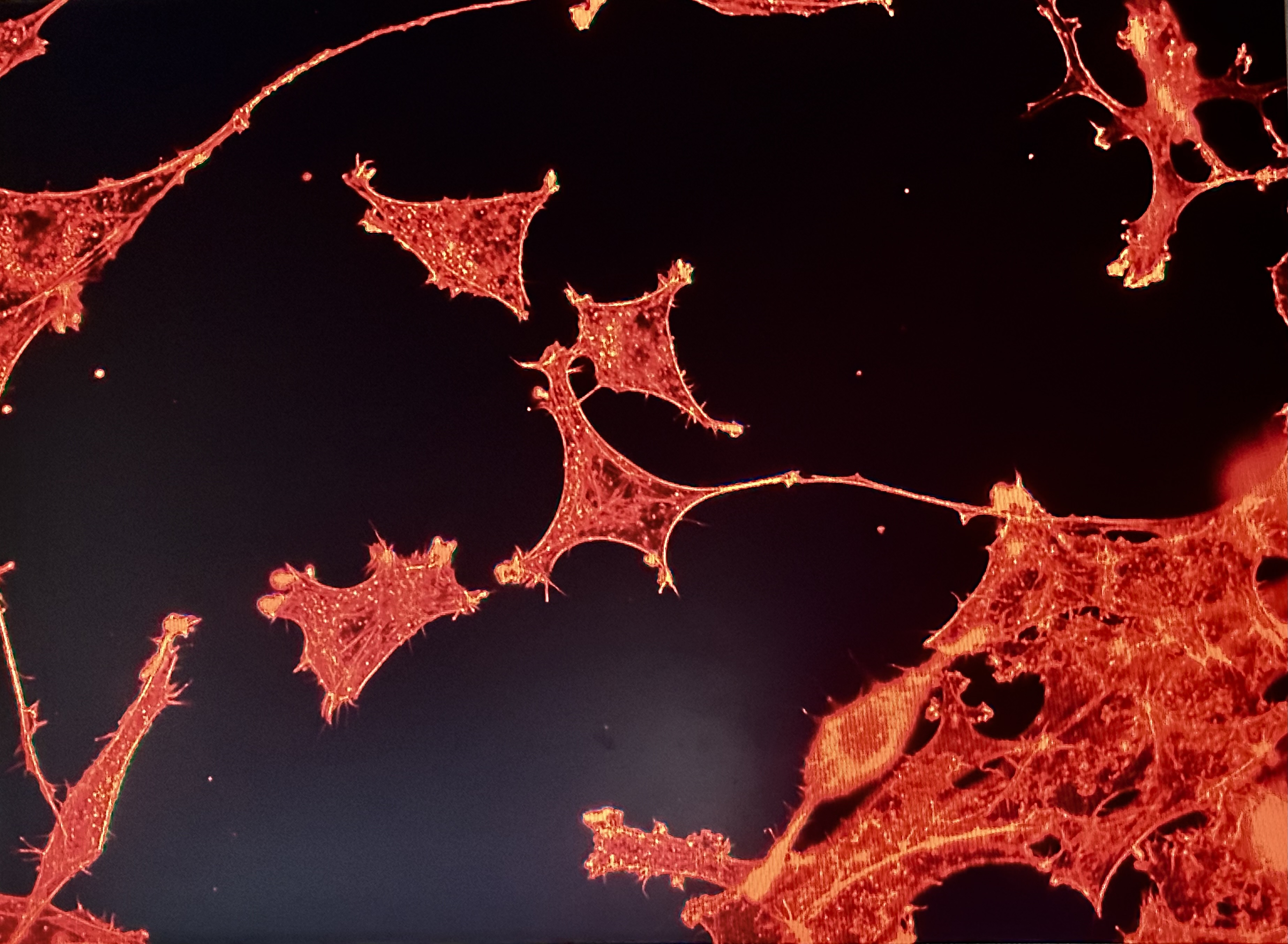
The MCF7 and MDAMB 231 cells were allowed to grow on circular coverslips in 24 well plates containing media. One set of cells(4 replicates) underwent serum starvation, another set of replicates were treated with TGF beta or TGF beta inhibitor for 24 hours. The experiment was also repeated for a duration of 48 hours for each of the cell lines. These coverslips were then washed with pbs thrice, they are then treated with triton for five minutes (to increases the permeability of the stains into the cells) and then washed with pbs again followed by treatment with phalloidin rhodamine(stain that binds to F-actin, this step is done in the dark) for one hour followed by three pbs washes and then treatment with DAPI(nuclear stain) for ten minutes. These stained coverslips are fixed onto glass slides and then visualised under the epifluorescence microscope. The actin filaments were visualised in red colour ( yellow excitation red emission) and the nuclei were visualised in blue(green excitation, blue emission). Around 25 photos of each replicate were taken using the microscope in order to assess the effects of the serum starvation, TGF beta and inhibitor treatment with respect to the controls on the surface area and the aspect ratios of the two types of cell lines. I also learnt staining the cells for antibody to vinculin. I further received an introduction into the operation of a confocal microscope which unlike the epifluorescence microscope was not limited to a 2 dimensional imaging of the cells but instead provided a more comprehensive 3 dimensional view of the cellular morphology at various z levels.
Results of the experiments
Literature review
The aggressiveness of Cancer cells are predominantly determined by their invasiveness and their ability to metastasise to distant sites. In the process of malignant transformation the cancer cells lose their dependence on integrin-mediated interactions with the extracellular matrix and genetic mutations cause a decrease in the expression of e cadherins which are crucial for cell to cell adhesion. There are also alterations in the hemidesmosomes that anchor the cells which disassemble causing integrity to interact with actin filaments which lead to an epithelial to mesenchymal transition of the cancer cells thereby rendering them more motile and invasive. The EMT is also associated with Cadherin switching from e to n cadherin subtype and the density of the extra cellular matrix has also shown to have an effect of collective cell migration in cancer cells. In addition the role of growth factors in cancer cell invasion has also been implicated with both n cadherin and FGFR and EGFR receptors being over expressed in cancer cells leading them to have increased motility and invasiveness.
This makes these integrins, cadherins and growth factors promising targets for the development of novel anti cancer therapeutic drugs.[2] Metastasis of cancers to distant sites from the primary tumour is a process governed by multiple steps: (1) Local tumor invades past the basement membrane into the surrounding stroma, (2) entry into the vasculature, (3) survival of the cells in the circulatory system, (4) extravasation of the cells into the parenchyma of the distant organ, (5) microscopic colonization of the distant organ, and (6) re-initiation of proliferation forming macroscopic metastases. Phenotypic plasticity controlled by various epigenetic mechanisms of the cancer cells enable them to adapt to the ever changing environmental conditions to ensure successful metastasis. Cancer cells that lack interactions with neighboring cancer cells can detach from the primary mass and migrate individually. The two different mechanisms of single-cell invasion are amoeboid and mesenchymal migration and is governed by the rigidity of the cell-matrix adhesions, the capacity of the tumour cells to remodel the ECM, and the contractility of the cytoskeleton. Collective cell migration on the other hand involve cancer cells that retain intact cell–cell adhesions while invading the tumor microenvironment and often encompass a ‘leader cell’ with altered polarity at the invasive margin which act as a guide for the other ‘following’ cancer cells with intact polarity that are attached to the leaders rear end and migrate taking advantage of the pulling force exerted by the leader cells.
A detailed study of the intrinsic processes involved in this collective cancer cell migration would also open new avenues for cancer chemotherapy targets. [3] The greatest challenge in developing anti cancer chemotherapy is the high levels of heterogeneity depicted by cancer cells. Within a single tumour there are usually present several micro environments of genetically heterogeneous cancer cells with varying levels of invasive and metastatic potential and varying levels of cellular differentiation. The heterogeneity in terms of critical cellular adhesive strength to the substrates could be determined by using a fluid shear device to detach cancer cells from the substrate and looking at the time required for each type of cell line(MDAMB 231 and MCF7) to detach completely. Collecting the detached cells at different time intervals during the process and subjecting them to genetic analysis via PCR helps identify the overexpression of genetic regions specific in determining increasing cell motility and invasiveness. Traction force microscopy experiments also show greater traction exerted by the more invasive cell lines.[1] As elucidated earlier cancer cells often show an overexpression of growth factor receptors like FGFR and EGFR.
TGF-β is yet another multi-functional cytokine that has been implicated in cancer cell invasion and metastasis. The cellular response to TGF-β is context dependent and can be either tumor suppressive or promoting based on the regulation of target genes and can lead to EMT transition and cadherin switching from e cadherin to n cadherin. TGF beta is often upregulated in breast cancer cells and is implicated in alteration of the cytoskeletal organization through increased expression of α-SMA causing the breast cancer cells to switch to a more migratory phenotype. Hence an earlier study aimed to quantify the effects of TGF beta on the bio mechanical properties of breast cancer cells. Highly invasive MDAMB 231 and less invasive MCF-7 breast cancer cells were subjected to 24 hours treatment with TGF beta following which their elastic and visco elastic properties were measured by atomic force microscopy using a spherical probe. The invasive MDAMB 231 cells showed increased fluid like nature with decreased moduli of elasticity as compare to MCF-7 which showed a stiffening response. The response at 48 hours remained similar in the MDAMB 231 cells with increase in elasticity while the MCF-7 cells showed a delayed response with increase in viscosity no relaxation times after 48 hours of TGF beta treatment. [4] Our project aimed to investigate further the effects of TGF beta and TGF beta inhibitor on the deadhesion of the cancer cells when subjected to the fluid shear stress device and the morphology of the cells in terms of the surface area and the aspect ratio of the cells which could provide further insights into the effects of TGF beta on the metastatic potential of breast cancer cells.
References
(1) Paddillaya N, Ingale K, Gaikwad C, Saini DK, Pullarkat P, Kondaiah P, Menon GI, Gundiah N. Cell adhesion strength and tractions are mechano-diagnostic features of cellular invasiveness. Soft Matter. 2022 Jun 15;18(23):4378-4388. doi: 10.1039/d2sm00015f. PMID: 35611829.
(2) Janiszewska M, Primi MC, Izard T. Cell adhesion in cancer: Beyond the migration of single cells. J Biol Chem. 2020 Feb 21;295(8):2495-2505. doi: 10.1074/jbc.REV119.007759. Epub 2020 Jan 14. PMID: 31937589; PMCID: PMC7039572.
(3) Teeuwssen M, Fodde R. Cell Heterogeneity and Phenotypic Plasticity in Metastasis Formation: The Case of Colon Cancer. Cancers (Basel). 2019 Sep 14;11(9):1368. doi: 10.3390/cancers11091368. Erratum in: Cancers (Basel). 2020 May 28;12(6): PMID: 31540068; PMCID: PMC6770401.
(4) Kulkarni AH, Chatterjee A, Kondaiah P, Gundiah N. TGF-β induces changes in breast cancer cell deformability. Phys Biol. 2018 Aug 14;15(6):065005. doi: 10.1088/1478-3975/aac3ba. PMID: 29745937.